








| 查看: 3080 | 回复: 12 | |||
| 当前只显示满足指定条件的回帖,点击这里查看本话题的所有回帖 | |||
[求助]
Image J 统计孔尺寸 已有2人参与
|
|||

请问大神们,Image J 软件怎么计算图片中的泡孔数目和尺寸????万分感谢!!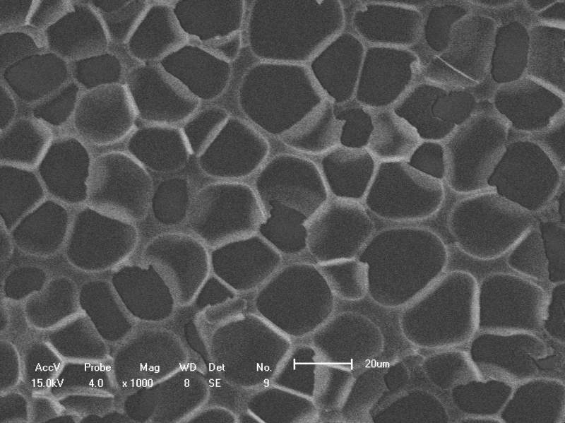 6-2.jpg |
 回复此楼
回复此楼» 收录本帖的淘帖专辑推荐
 发泡 发泡 |
» 猜你喜欢
 售SCI一区文章,我:8.O.55.1.O.54,科目齐全,可伽急
已经有5人回复
售SCI一区文章,我:8.O.55.1.O.54,科目齐全,可伽急
已经有5人回复
-
有多少人是今天查系统知道结果的?
已经有18人回复
-
售SCI-T0P文章,我:8O.5.5.1.O.54,科目齐全,可+急
已经有5人回复
-
售SCI文章,我:8O.5.5.1O.54,科目全,可十急
已经有4人回复
-
售SCI一区文章,我:8.O.55.1.O.54,科目齐全,可伽急
已经有5人回复
-
我就是申请一个面上项目而已,这评审意见是按照杰青的条件评的吧?
已经有4人回复
-
国自然评审意见
已经有15人回复
-
为什么资助数各大高校都创新高,自己申请怎么就这么难
已经有13人回复
-
国自然面上复盘~欢迎讨论
已经有14人回复
-
系统查不到
已经有11人回复
dota
木虫 (著名写手)
- 应助: 19 (小学生)
- 金币: 1743.5
- 散金: 3711
- 红花: 9
- 帖子: 1258
- 在线: 1385.2小时
- 虫号: 302890
- 注册: 2006-12-03
- 性别: GG
- 专业: Chem & Biomed Imaging

5楼2015-11-24 11:04:46
dota
木虫 (著名写手)
- 应助: 19 (小学生)
- 金币: 1743.5
- 散金: 3711
- 红花: 9
- 帖子: 1258
- 在线: 1385.2小时
- 虫号: 302890
- 注册: 2006-12-03
- 性别: GG
- 专业: Chem & Biomed Imaging
【答案】应助回帖
感谢参与,应助指数 +1
|
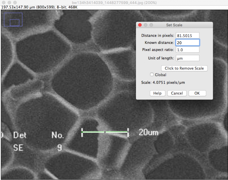
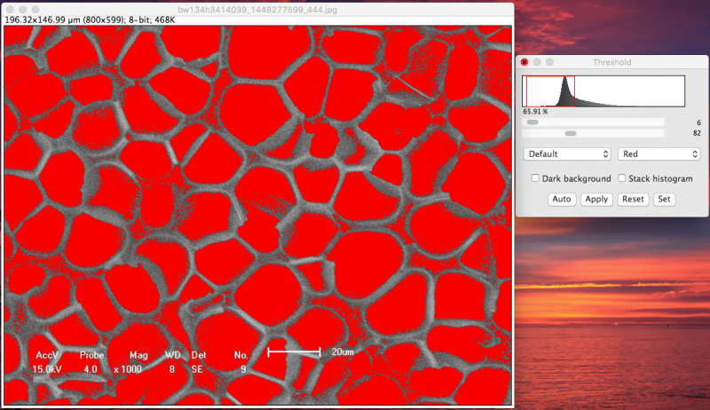
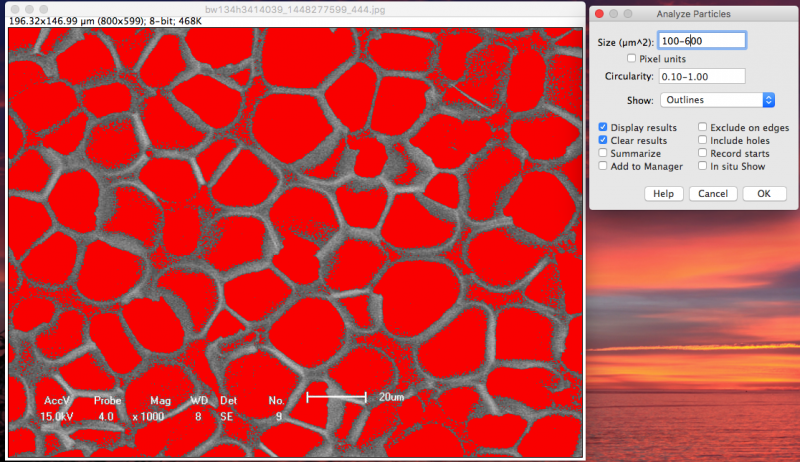
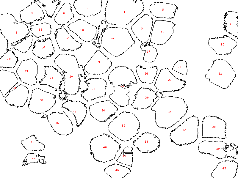
1) Set scale. Enlarge your image, draw a line of the exactly same length to the scale bar. For you image, 81.5 pixel = 20 um) 2) Adjust threshold, which showed reasonable segmentations. 3) Analysis Particles. Based on the visional pore sizes, I would say areas between 100 um^2 and 600 um^2 should be the correct pores. 4) Copy the results into the Excel to calculate the average and the stDev. The 45 pores were detected out. The pore perimeter is 25.6 +/- 9.8 um. Some pore might be missed due to either the irregular shapes or the low image contrasts (not good segmentations) Cheers! |
2楼2015-11-24 09:36:04
dota
木虫 (著名写手)
- 应助: 19 (小学生)
- 金币: 1743.5
- 散金: 3711
- 红花: 9
- 帖子: 1258
- 在线: 1385.2小时
- 虫号: 302890
- 注册: 2006-12-03
- 性别: GG
- 专业: Chem & Biomed Imaging
3楼2015-11-24 09:38:56
 太感谢了!!还有一个问题,就是阈值要怎么调才算合适?Auto 好像不太对
 4楼2015-11-24 10:12:29


 10
10